IMAGING SOLUTIONS > MOBILE SOLUTIONS
Unmatched Precision On-The-Go with Mobile Imaging Solutions
In today’s dynamic healthcare landscape, the need for precise diagnostic imaging doesn’t stop at the provider’s door. At COL Imaging, our mobile imaging solutions redefine this unexplored frontier and bring unmatched precision directly to patients. Whether it’s a rural area, a busy metropolitan hospital, outpatient imaging center, or physician clinic setting, we ensure that top-tier diagnostic accuracy is always within reach. Experience the future of medical imaging with our unparalleled ready-to-go MRI, CT, and X-Ray solutions, where every scan is a testament to clarity, convenience, and care.
Taking Advanced Diagnostics On The Road
In an era where medical innovation converges with budgetary restraints, it’s important to find the right imaging solutions for your needs. Mobile imaging solutions bridge the gap between advanced diagnostic imaging and the diverse needs of populations – both urban and rural. By mobilizing top-tier diagnostic tools, these solutions transcend traditional clinic boundaries, delivering unparalleled precision whenever and wherever it’s needed. Now, healthcare providers can ensure that no patient is left without access to the best diagnostic care, regardless of location, with mobile diagnostic solutions from COL Imaging Solutions.
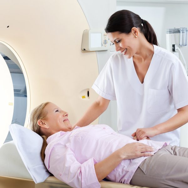
At the intersection where MRI technology meets mobility, COL Imaging’s mobile MRI solutions ensure top-tier diagnostics are accessible beyond the confines of a traditional setting. As leaders in innovative imaging healthcare solutions, we have seamlessly integrated the depth and precision of MRI scanning into a mobile platform, allowing us to bring affordable and quality diagnostic clarity directly to your doorstep. Dive into a transformative approach to diagnostic imaging, where every journey echoes our commitment to flexibility, accessibility, affordability, and credibility.
COL Imaging’s mobile CT solutions bring superior diagnostics out of the traditional clinical setting. We’ve successfully merged the detailed accuracy of CT scans with a mobile unit. This enables us to deliver unmatched diagnostic clarity to any location. Experience a revolutionary method of patient care with us, where each step reflects our commitment to quality service, accessibility, flexibility and affordability.
Frequently Asked Questions
Mobile imaging is a subset of telemedicine, referring to the use of portable imaging devices or technologies to obtain diagnostic scans outside of a traditional fixed-location radiologist or imaging department. This kind of imaging is especially useful when traditional imaging facilities are not easily accessible or when patients are immobile.
A mobile imaging unit is typically an 8’ x 48’ customized specialty medical trailer that has been specially designed and equipped with advanced diagnostic imaging equipment. The purpose of these units is to bring diagnostic imaging capabilities to locations where they might not be readily available – such as in rural or underserved areas, facilities with limited available space – coverage for equipment downtime or to provide temporary imaging service in places experiencing high demand.
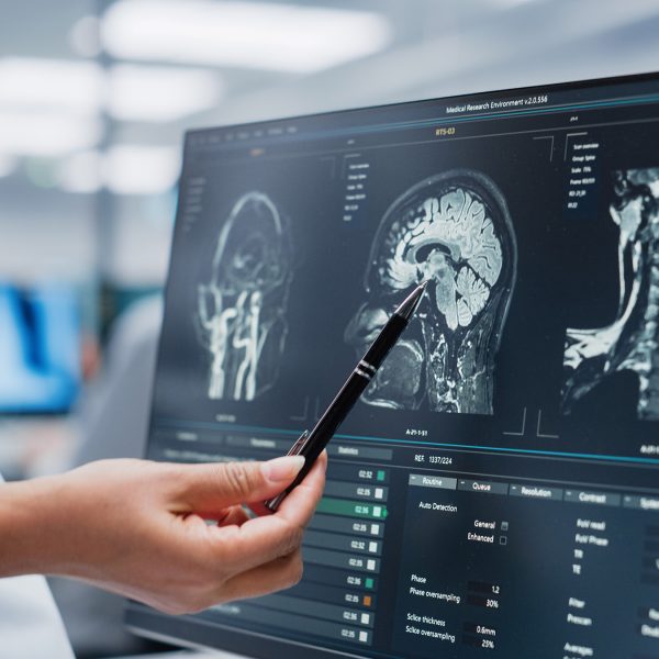
Digital imaging has revolutionized how healthcare professionals visualize, analyze, and interpret physiological and pathological conditions. There are several techniques and machines designed to perform different types of imaging, including computed tomography (CT or CAT scans), magnetic resonance imaging (MRI), positron emission tomography (PET scans), ultrasound, and more. Which imaging modality will be used is determined by a combination of clinical indications, what specific information the physician needs, the patient’s medical history, and available resources.
Mobile imaging offers several advantages to both physicians and patients. Mobile imaging units can reach populations in rural or underserved areas where traditional imaging facilities might not be available. In emergencies, such as natural disasters, mobile units can be dispatched to affected areas to provide critical diagnostic services on the spot. For areas where it may not be economically feasible to set up a permanent facility, mobile imaging units can offer the cost-effective alternative.
MRI, or Magnetic Resonance Imaging, is a widely used imaging technique that offers detailed views of internal structures without ionizing radiation. However, despite its general safety, there are potential side effects and considerations. Some MRI exams utilize a gadolinium-based contrast agent to enhance image clarity. Though rare, allergic reactions may occur that range from minor skin reactions to more severe respiratory issues. A rare condition called Nephrogenic Systemic Fibrosis has been linked to gadolinium-based contrast dye in patients with significant kidney issues. The machine itself produces loud noises, which some patients find uncomfortable even with earplugs or headphones. Additionally, the confined space inside the MRI scanner can induce feelings of claustrophobia in certain patients. Moreover, the MRI’s strong magnetic field can interact with metal implants or fragments in the body, which poses a significant potential risk. Patients with metal implants such as stents, filters, pacemakers, joint replacements, screws, metallic fragments like bullets or shrapnel, or other metallic devices cannot be considered for an MRI due to this inherent risk.
An MRI (Magnetic Resonance Imaging) and a CT (Computed Tomography) scan are both advanced diagnostic imaging techniques, but each is suited for distinct purposes. An MRI employs powerful magnets and radio waves to generate detailed images of soft tissues without ionizing radiation, making it especially effective for visualizing the brain, spinal cord, joints, and muscle tissues.
A CT scan uses a series of X-rays from various angles to create cross-sectional images of the body. CT scans are particularly valuable for visualizing bones, lungs, and certain soft tissues. A CT scan can capture images quickly, which is beneficial in emergency situations. Because CT scans use ionizing radiation, there’s a radiation exposure risk. The choice between these modalities depends on your physician’s specific clinical questions and the type of tissue being examined.
An X-ray and a CT (Computed Tomography) scan both use ionizing radiation to capture images of the body’s internal structures, but they operate on different principles and serve different diagnostic purposes. Traditional X-rays produce two-dimensional images by passing a controlled amount of radiation through the body, capturing images on a film or digital sensor. They are often used for viewing bones, detecting fractures, and checking for lung infections.
In contrast, a CT scan uses a rotating X-ray machine to take multiple images from different angles. A computer processes these images to produce cross-sectional, and often three-dimensional, views of the area. This allows for more detailed visualization, particularly of soft tissues, organs, and blood vessels. While both X-rays and CT scans are valuable diagnostic tools, CT scans provide a more comprehensive view but come with a higher dose of radiation compared to standard X-rays.